Producing a Great Stifle Image
- Kevin Scholz
Though I am NOT a DVM, I hang around you guys way too much.... Last month I had an opportunity to spend a day with a veterinary friend/client in southern Wisconsin. We spent part of the day taking x-rays of several equine joints. The goal - test and document some of the potential combinations of x-ray generator and plate positioning, x-ray technique, and Metron image processing to produce the maximum diagnostic quality x-ray images.
With CR x-ray, you have more flexibility with x-ray technique and image processing. We have the option to increase x-ray power (kVp & MAS) adding more data to the electronic image but we need to correspondingly process the image so it does not appear over-exposed. Yes, some x-ray shots demand more power than the standard portable x-ray generator (kVp=80, MA=15) can produce - not always powering enough energy through the object of interest to present good detail on the plate.
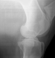
The Stifle presented the biggest challenge to producing a diagnostic image. Yet, the Stifle merely demanded an exaggeration of the same methods used in other x-ray shots. With the Stifle, we positioned much closer with a focal distance of 16 inches and set the focal point about 2 inches above the joint. We also pointed down at about 15% to help match the angle position of the plate held to the opposite side.
Using a time of 0.18 seconds (a bit higher that most might be comfortable with) we produced an image with great detail throughout. The other Equine joints do not need the close focal distance or exaggerated time to produce great images, but we did still push beyond what has been "normal" for x-ray exposure.
Visit the new Metron
"Images and Videos" page to see more examples of Equine images impacted by improvements in the process of image acquisition. Images presented include x-ray technique, focal length, and veterinary practice source.


 It was great to talk with and meet many of you at the AAEP show in Las Vegas.
 Dr Fox has been using Metron Imaging Software and Support Services since October 2009. In addition to the standard installation, set up, and online education, Dr Fox signed up for our VetRay conversion service to convert his old images to Metron "veterinary" DICOM standard. This allows him to keep all the old and new images together in the Metron Imaging Software.